

Xcalibur Diffractometer
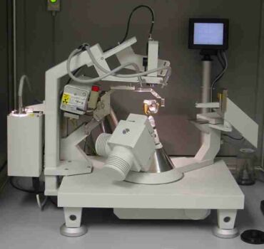
This is the Xcalibur diffractometer from Oxford Diffraction
It is a kappa geometry diffractometer, which means it does not have the large Eulerian cradle of a conventional 4-circle diffractometer, such as our Huber. Instead it is equipped with a "kappa" arm which can be seen in the picture pointing towards you. The kappa arm rotates on the omega-axis and carries the phi-axis.
The advantages of the kappa geometry include open access to the sample, and the ability to build an unrestricted small goniometer; intensities are therefore higher than from a larger goniometer. This makes it ideal for precise intensity data collections from single-crystals held at high-pressures in diamond-anvil cells. These data will allow us to determine how the distances and angles between atoms change as the material is subject to pressure.
The diffractometer is equipped with both a point detector system and a Sapphire-II CCD camera that can be mounted simultaneously.
See the page about structures at high pressure for a more detailed description of the methods we use, or read the recent MSA Reviews in Mineralogy volume 41 on High-Pressure and High-Temperature Diffraction.
The Xcalibur diffractometer system was delivered in June 2001. Here are some pictures from the installation.
For further information please contact the instrument scientist Dr. Jing Zhao (Tel: 231-5539).
In the late 1970's the International Union of Crystallography distributed a set of high-quality crystals of ruby ground into spheres of 150-200 microns on diameter. These crystals have become a standard test of data collection procedures on single-crystal X-ray diffractometers.
We collected several datasets from our ruby sphere with various step sizes and selections of slits to define different detector apertures on the Xcalibur.
We found that a 1 degree-wide horizontal detector aperture was too narrow and resulted in the clipping of diffraction profiles at higher angles. Wider slits resulted in peak profiles with a FWHM of 0.16degrees in omega.
With the generator running at 40KV/30mA data collections of a hemisphere of reflections out to 80deg 2theta (909 reflections) took about 21 hours (details below).
It is a kappa geometry diffractometer, which means it does not have the large Eulerian cradle of a conventional 4-circle diffractometer, such as our Huber. Instead it is equipped with a "kappa" arm which can be seen in the picture pointing towards you. The kappa arm rotates on the omega-axis and carries the phi-axis.
The advantages of the kappa geometry include open access to the sample, and the ability to build an unrestricted small goniometer; intensities are therefore higher than from a larger goniometer. This makes it ideal for precise intensity data collections from single-crystals held at high-pressures in diamond-anvil cells. These data will allow us to determine how the distances and angles between atoms change as the material is subject to pressure.
The diffractometer is equipped with both a point detector system and a Sapphire-II CCD camera that can be mounted simultaneously.
See the page about structures at high pressure for a more detailed description of the methods we use, or read the recent MSA Reviews in Mineralogy volume 41 on High-Pressure and High-Temperature Diffraction.
The Xcalibur diffractometer system was delivered in June 2001. Here are some pictures from the installation.
For further information please contact the instrument scientist Dr. Jing Zhao (Tel: 231-5539).
Ruby test datasets
In the late 1970's the International Union of Crystallography distributed a set of high-quality crystals of ruby ground into spheres of 150-200 microns on diameter. These crystals have become a standard test of data collection procedures on single-crystal X-ray diffractometers.
We collected several datasets from our ruby sphere with various step sizes and selections of slits to define different detector apertures on the Xcalibur.
We found that a 1 degree-wide horizontal detector aperture was too narrow and resulted in the clipping of diffraction profiles at higher angles. Wider slits resulted in peak profiles with a FWHM of 0.16degrees in omega.
With the generator running at 40KV/30mA data collections of a hemisphere of reflections out to 80deg 2theta (909 reflections) took about 21 hours (details below).
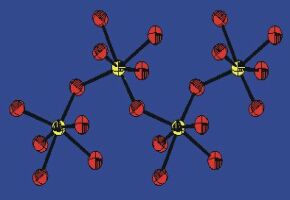 |
This is a thermal ellipsoid plot of the structure of ruby refined to the Xcalibur data.
Note that the ellipsoids of the aluminium atoms are almost spherical, while those of the oxygen atoms show the smallest radius (i.e. smallest thermal vibration) along the Al-O bonds. In fact, these ellipsoids and the displacement parameters are indicative of rigid-body behaviour of the AlO6 octahedra to within 2%, a good indication of the high-quality of the data and structure refinement. |
||||||||||||||||||||||||||||
|
Data collection details.
Omega scans, 60 steps, width=1.2deg, initial speed 0.05 deg/minute
Constant precision mode: to 33 I/sigma, initial rejection level 0.5 I/sigma. Minimum speed 0.005 deg/minute |
Data reduction details. |
||||||||||||||||||||||||||||
|
Refinement. with RFINE-99 (see software)
10 refined parameters (positions, aniso displacement, scale factor and isotropic extinction) Ru = 0.013, Rw = 0.015, Gfit = 0.95 on 176* obs * In all datasets we found the 006 reflection to have Fobs>>Fcalc. We suspect that this is the result of lambda/2 diffraction from the 00 12 reflection. The 006 reflection was therefore excluded from all refinements. |
Results
Al-O = 1.9719(3) and 1.85500(15) A |
Lessons Learned (from duplicate refinements)
The quality of the refinement for such a strong scatterer is not significantly affected by the choice of detector slit sizes, but atomic displacement parameters do increase by about 10% if the slits are too small.
The choice of step size in the scans makes little difference. The internal consistency of datasets, as measured by Rint, collected with the Xcalibur system is amazingly good: the Gfit values, all < 1 indicate that the estimated uncertainties in reflection intensities are actually over-estimated even using Blessing's averaging criteria.
The choice of step size in the scans makes little difference. The internal consistency of datasets, as measured by Rint, collected with the Xcalibur system is amazingly good: the Gfit values, all < 1 indicate that the estimated uncertainties in reflection intensities are actually over-estimated even using Blessing's averaging criteria.
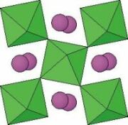
CaSnO3 perovskite
| CaSnO3 perovskite provides a good test of the quality of data collection, reduction, and structure refinement procedures because it has a moderately high absorption coefficient for Mo radiation (~122cm-1), orthorhombic symmetry, and good-quality crystals can be grown (see Kung et al. 2000 in my publications). | We collected a test intensity dataset from CaSnO3 on the Xcalibur system:
Crystal size: 120x120x130 micron 2theta_max =80 degrees, 4 asymmetric units, 2919 reflections. Steps-cans, 1.2deg wide, 60 steps, constant precision mode to I/sigma = 10. Min speed 0.005deg/min. Generator at 50KV/40mA Data collection time was 84 hours. |
Data reduction
Integration by profile fitting.
Absorption by ABSORB (Burnham, 1966) Averaging in mmm: Rint = 0.008 on 754 averaged reflections with F > 4sigF Refinement: Ru=0.019, Rw=0.026, Gfit=1.7 on 762 Fobs, 29 parameters |
 |
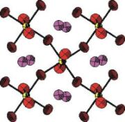
Results
The three independent Sn-O bonds within the SnO6 octahedra show only a slight distortion:
2.0532(8), 2.0539(4), and 2.0572(8) A and a small angular distortion of less than 2 degrees, despite a large tilt angle of 148 degrees. The anisotropic displacement parameters (right) are consistent with rigid-body motion of the SnO6 octahedra: note how the ellipsoids of the oxygen atoms are elongated perpendicular to the Sn-O bonds. It is also clear that the O atom motion is not influenced by the Ca atoms |
 |