 Two new crystal forms of
Calmodulin from Chicken Two new crystal forms of
Calmodulin from Chicken
Two new crystal forms of
Calmodulin from Chicken Two new crystal forms of
Calmodulin from Chicken
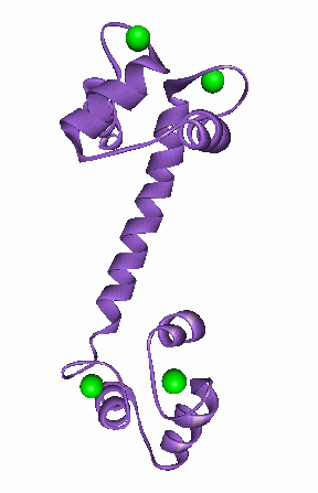
Calmodulin (CAM) is a highly conserved eukaryotric protein that plays an important role
in Ca-dependent signal transduction pathways. Its structure was first solved at 3.0 Ĺ
resolution using multiple isomorphous derivatives of rat testis CAM. Structures of various
other CAMs have since been reported: bovine brain at 2.2 Ĺ, recombinant Drosophila
melanogaster and recombinant Paramecium tetraurelia. All these studies reported triclinic
crystals by precipitation with 2-methyl 2,4-pentanediol (MPD), with resolution limits
between 3.0 and 1.8 Ĺ. In these P1 crystals, CAM adopts a dumbell shape with two lobes connected by a central 21
residue helix. NMR studies in solution, indicate a flexible region in this helix allowing
it to fold around target peptides. A mutant CAM (des-Glu84) provides evidence in
support of this flexible tether. Since all previously reported native CAMs are essentially
isostructural in P1, investigation of other crystal forms is important.
 Crystallization of P1
crystals
Crystallization of P1
crystals
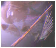
P1 crystals of CAM were difficult to grow by reported methods. The procedure of
Cook & Sack gave thin needles diffracting to ~2.5 Ĺ, but were unsuitable for data
collection. The procedure was modified as follows. Crystals grew at room temperature
within 2 days in 10 ml hanging drops (6 ml CAM (12.5 mg/ml in 4 mM CaCl2), 4 ml reservoir
(10 mM NaOAc, pH 4.0, 25% MPD, 15% iso-propanol)). Small, well-formed seed crystals were
washed in a different reservoir (10 mM NaOAc, pH 4.0, 30% MPD, 15% iso-propanol), and
placed in drops of 12 ml CAM and 8 ml of reservoir. Some crystals grew large within a few
days, but then stopped growing. These lancet-like crystals (~2 x 0.2 x 0.1 mm), were
fragile and delaminated into flat shreds parallel to the long growth direction.
Crystallization of P212121 crystals
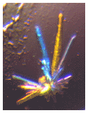 A mini-screen containing a subset of the Hampton Research Crystal Screen I was set up
to determine alternate crystallization conditions. Only condition 31 (30% PEG 4000, no
buffer, 0.2 M (NH4)2SO4(aq)) delivered crystals, but these were tiny and embedded in a
cloudy precipitate. A 4 by 6 screen of hanging drops was set up at room temperature as
follows: 4 ml of CAM (15 mg/ml in 4 mM CaCl2) mixed with 2.7 ml of reservoir containing 10
mM of NaOAc (pH 3.7-4.4) and 5 to 20 % PEG 4000, pH in the wells was not adjusted. Within
two days, prismatic crystal clusters appeared at PEG concentrations of 15% and 20% in the
pH range 3.85 to 4.14 (i.e. around the isoelectric point).
A mini-screen containing a subset of the Hampton Research Crystal Screen I was set up
to determine alternate crystallization conditions. Only condition 31 (30% PEG 4000, no
buffer, 0.2 M (NH4)2SO4(aq)) delivered crystals, but these were tiny and embedded in a
cloudy precipitate. A 4 by 6 screen of hanging drops was set up at room temperature as
follows: 4 ml of CAM (15 mg/ml in 4 mM CaCl2) mixed with 2.7 ml of reservoir containing 10
mM of NaOAc (pH 3.7-4.4) and 5 to 20 % PEG 4000, pH in the wells was not adjusted. Within
two days, prismatic crystal clusters appeared at PEG concentrations of 15% and 20% in the
pH range 3.85 to 4.14 (i.e. around the isoelectric point).
Preliminary x-ray analysis of P1 crystals
The MPD concentration in the mother liquor was sufficient to form a protective glass upon
cooling. Crystals of varying quality were mounted in fine glass loops embedded in copper
mounting pins, and quickly transferred to the (initially deflected) cold-stream of a
modified Siemens LT-2 low-temperature apparatus. The crystals had wide (~2°
base-to-base), multiply peaked spot profiles. Eventually, a 1.0 x 0.2 x 0.1 mm fragment
broken from a large aggregate gave single-peaked diffraction maxima that were ~1.5°
base-to-base and ~0.6° full-width-at-half-maximum (FWHM) in w. The tendency to splinter
is a likely cause of the wide, multiply peaked diffraction spot profiles.
The crystals were triclinic (a = 59.7 Ĺ, b = 53.1 Ĺ, c = 24.6 Ĺ, a = 93.2 °, b =
96.7° , g = 89.2°, Z = 2), and appear to be related to other P1 CAM crystals with Z=1,
but with the a-axis doubled. On density considerations, solvent content in these 'cell
doubled' P1 crystals is estimated at about 50% by volume. A dataset to 1.8 Ĺ was
collected at 130 K on a Xuong-Hamlin area detector.
The structure has been solved by molecular replacement (Trakhanov and Rupp) and has been
refined.
Preliminary x-ray analysis of P212121 crystals
These crystals could not be mounted in their mother liquor. After surgery to break the
clusters, cooling was successful after a brief wash in unbuffered aqueous 50% PEG 4000.
Visual inspection after some minutes suggested that little could be gained by longer
soaks. The crystals were mounted in glass loops as described above. All gave sharp,
single-peaked diffraction spots with base-to-base and FWHM profiles of ~0.8° and ~0.3°
respectively. They diffracted well to around 1.4 Ĺ, and in all cases, some appreciable
intensities were observed above 1.2 Ĺ. A crystal approximately 0.30 x 0.15 x 0.10 mm was
indexed as orthorhombic, space group P212121 with a = 32.2 Ĺ, b = 56.0 Ĺ, c = 67.3 Ĺ, Z
= 4. On density considerations, solvent content is about 30%, much lower than the
triclinic form. This may be linked to the dramatically higher resolution, and hence to a
more ordered structure. Data extending to 1.2 Ĺ were collected at 130 K in separate low
and high resolution shells, statistics are given in table 2. Better counting statistics at
high resolution should be possible when crystallization conditions have been further
optimized. In the combined set (Rmerge = 4.12%, <I>/<s(I)> = 11.5),
completeness to 1.7 Ĺ is 83.6%, while at the highest resolution it is ~40 %.
Diffractometer modifications made since these experiments will allow recording of a full
high-resolution dataset. The structure is currently under refinement.
Contributing Scientists: Sean Parkin and Bernhard Rupp, LLNL - BBRP: Crystallography, Dan Marshak, Cold Spring Harbor Laboratory : Showed us how to prepare Calmodulin, Mark Knapp (LLNL) : refinement of crystal growth conditions
Initial results published in : Crystallization And Preliminary X-Ray Analysis Of Two New Crystal Forms Of Calmodulin, B.Rupp, D.Marshak and S.Parkin, Acta Crystallogr. D 52, 411 (1996)
Click the file names to access a PDB entry of Calmodulin (1clx15.pdb)
If you need a viewer to look at the PDB structures, try RasMol - also for Windows (RasWin).
![]() Back to The Macromolecular Crystallography Home Page
Back to The Macromolecular Crystallography Home Page
LLNL Disclaimer
This World Wide Web site conceived and maintained by
Bernhard Rupp (br@llnl.gov)
Last revised August 03, 2000 23:26
UCRL-MI-125269