

Diffuse scattering
When the atoms are completely ordered in a crystalline structure, then one only observes
sharp "Bragg" peaks in the diffraction pattern. When the atoms are not fully ordered, then the
intensity of the Bragg peaks is diminished and some of the scattered intensity appears as "diffuse"
scattering.
Professor Ulli Bismayer (from Hamburg) and Ross Angel have been working on a series of materials with the palmierite structure which display diffuse scattering as a result of a phase transition.
These data were also collected on our
Xcalibur-2 instrument, but with a 0.3mm Enhance optic shortened
to accomodate the diamond-anvil pressure cell. An ETH design of diamond-anvil cell equipped with
thin diamonds and steel seats was used in this experiment.Further details are
provided in an Acta Cryst. paper.
Professor Ulli Bismayer (from Hamburg) and Ross Angel have been working on a series of materials with the palmierite structure which display diffuse scattering as a result of a phase transition.
 |
This first set of three images are reconstructed sections through the diffraction
pattern of Pb3(P0.48As0.52)2O4 at room temperature and pressure. The data were collected on our
Xcalibur-2 instrument with a standard 0.8mm Enhance optic and a Sapphire-II CCD camera.
This material has a trigonal unit cell at room temperature as a result of a phase transition from monoclinic symmetry at lower temperature. This hk0 section clearly shows the 3-fold symmetry, and the strong Bragg reflections. You can also see sets of four or six weaker diffuse reflections around each Bragg reflection. |
 |
If the structure were truely trigonal on a local scale then this reciprocal lattice section at hk-1/2 would have no intensity. Instead we see some of the extra reflections that appeared as satellites around the Bragg reflections in the hk0 section. |
 |
The other weak satellites appear in this hk+1/2 section. All of these weak and diffuse reflections arise from the presence of small static clusters of atoms that retain the monoclinic distortion even while the crystal as a whole exhibits trigonal symmetry. |
| Pure lead phosphate undergoes the transition from monoclinic symmetry to
trigonal symmetry at a pressure of 1.8 GPa at room temperature. The three images on the right
are small parts of the reciprocal lattice sections collected at 2.0 GPa, just on the trigonal
side of the phase transition. From the top they are hk-1/2, hk0, and hk+1/2.
In the top and bottom images you should be able to see some very weak and diffuse satellite reflections. These diffuse satellites are more obvious in the intensity profiles along the arrowed lines. The middle image shows the hk0 layer with the much stronger Bragg spots; 300 is in the middle. We believe that this may be the first observation of diffuse scattering with a laboratory instrument from a crystal held at high pressures in a diamond-anvil cell. | 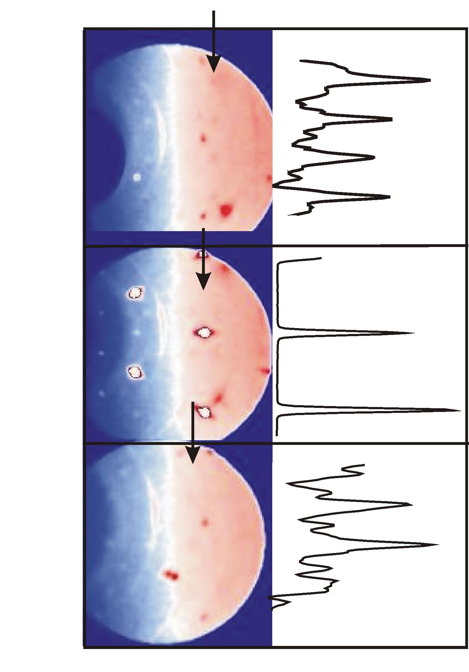 |