Crystal structure of a monoclonal 2E8 Fab antibody fragment specific to the low density lipoprotein receptor binding region ofApolipoprotein E refined at 1.9 Å
Sergei Trakhanov,1,2,3 Sean Parkin,3 Robert Raffai,4 Ross Milne4, Yvonne M Newhouse,1 Karl Weisgraber,1,2,5
and Bernhard Rupp3,*
1 Gladstone Institute of Cardiovascular Disease, 2 Cardiovascular Research Institute, 5Department of Pathology, University of California, San Francisco, CA 94141, USA
4 Lipoprotein and Atherosclerosis Group, University of Ottawa Heart Institute and Department of Pathology and Biochemistry, University of Ottawa, Ontario, K1Y 4E9, Canada
3 Biology and Biotechnology Research Program, Lawrence Livermore National Laboratory, Livermore, CA 94551, USA
Abstract - Introduction
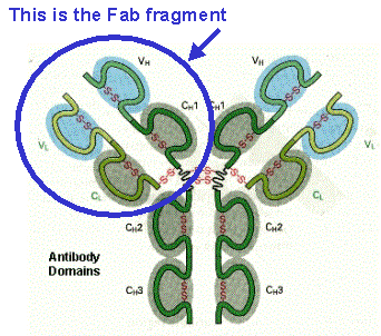
- Fab tutorial - Supplemental
Material - Published Paper - PDB entry 12E8
Antibody Search and Nomenclature
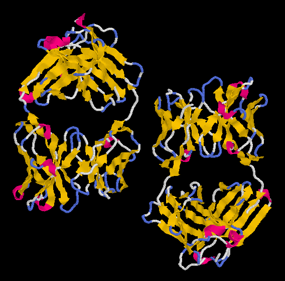
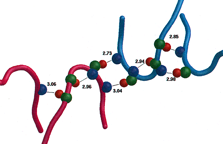
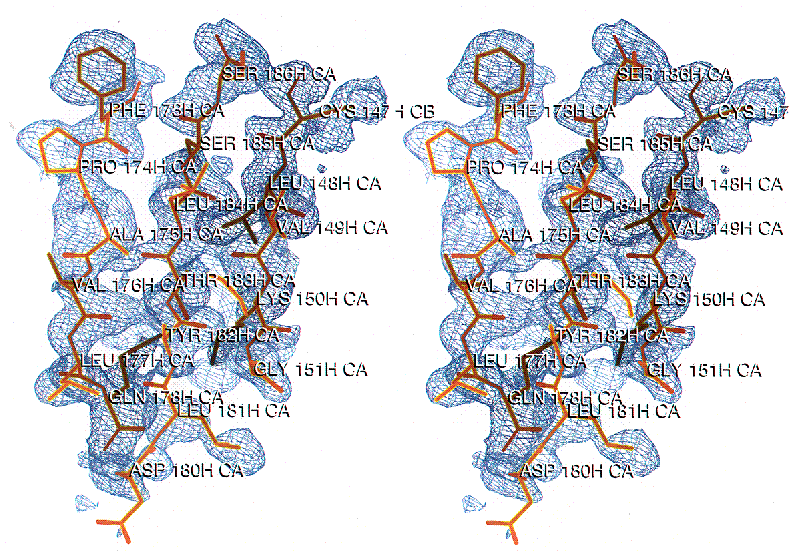
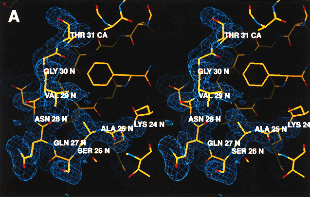
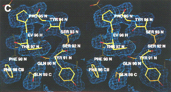
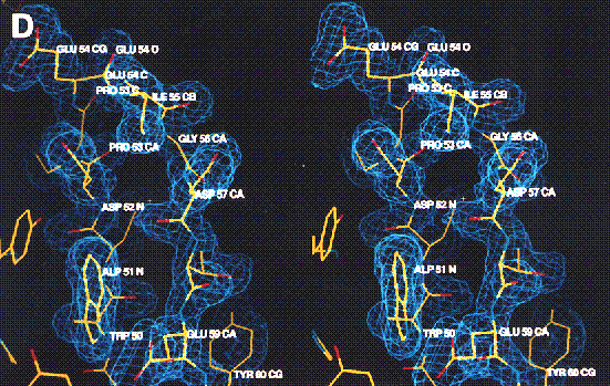
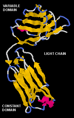
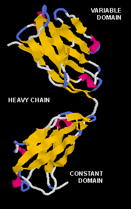
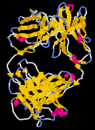
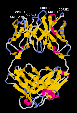
The crystal structure of the Fab fragment of 2E8, the monoclonal IgG1-kappa antibody specific to the low density lipoprotein (LDL) receptor binding region of apolipoprotein E (ApoE), has been solved by molecular replacement and refined at 1.95 Å resolution. Two 2E8 Fab molecules in the asymmetric unit are related by non-crystallographic symmetry, and are hydrogen bonded through a ß-sheet like intermolecular contact in the heavy chain complementarity determining region 3 (CDRH3) of each molecule. The structure has been refined to an R-factor of 0.21 (20 -1.95 Å). All CDRs are well defined . The frequently ill-defined heavy chain constant domain (CH) has been retraced with the aid of automatic refinement, confirming the ß-sheet tracing independently of any starting models. As a resolution better than 2 Å is not common for Fab fragments, this model represents a well-defined Fab structure and should prove useful in MR solution of other Fab fragments. Furthermore, in the absence of a LDL receptor structure, the homology of the 2E8 CDR2 to the ligand-binding domain of the LDL receptor has been exploited to model the ApoE – LDL receptor interaction.
Introduction (if you want a very basic introcuction go to More About Antibody Fragments)
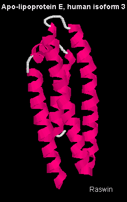
Apolipoprotein E (apoE, 34kD, 299 residues) is a protein component of several classes of human plasma lipoproteins (Mahley, 1988; Mahley et al., 1984; Weisgraber, 1994). As a ligand for the low density lipoprotein (LDL) receptor (see our recent work on defective receptor binding in E2) and other lipoprotein receptors in the LDL receptor family, apoE plays a major role in modulating plasma lipoprotein metabolism. Variant forms of apoE, including apoE2, possess defective binding to the LDL receptor and are an underlying cause of type III hyperlipoproteinemia, a disorder in lipid metabolism associated with premature heart disease (Mahley & Rall, 1995).
The structure of the apoE 22 kDa N-terminal fragment (residues 1-191) containing the LDL receptor binding region (residues 136–150) has been determined. (Wilson et al., 1991). The LDL receptor-binding region is characterized by a cluster of basic residues that are thought to interact with complementary clusters of acidic residues contained within the LDL receptor ligand-binding domain (Mahley, 1988). Detailed structural information on the LDL receptor is not yet available, although structures of cysteine-rich repeats 1 and 2 of the ligand-binding domain have been determined individually by nuclear magnetic resonance (NMR, Daly et al., 1995a; Daly et al., 1995b), and recently the structure of repeat 5 by x-ray crystallography (Fass et al, 1997).
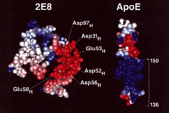
In the absence of complete structural information of the intact LDL receptor-binding domain, we sought an alternative approach to model the receptor:ligand interaction. Two monoclonal antibodies, designated 1D7 and 2E8, are known to block the interaction of apoE with the LDL receptor by binding to epitopes within the receptor-binding region of apoE (Raffai et al., 1995). Sequence alignment of the primary structures of the six complementarity-determining regions (CDRs) of the 1D7 and 2E8 antibodies with the seven cysteine-rich repeats of the LDL receptor reveals homology between the 2E8 heavy chain CDR2 and repeat 5 of the LDL receptor. Deletion mutagenesis analysis suggested that this repeat is involved in the interaction of apoE with the LDL receptor (Russell et al., 1989). Both 2E8 and the LDL receptor share a decreased affinity for apoE2, the isoform responsible for type-III hyperlipoproteinemia (Mahley & Rall, 1995). The 2E8 heavy chain CDR2 contains two glutamic acid residues and aspartic acid, which may mimic the interaction of apoE with the LDL receptor. Examination of that interaction should provide useful insights into the mechanism of apoE-LDL receptor binding. As a first step, we present the three-dimensional structure of the 2E8 Fab fragment at 1.95 Å resolution and initial modeling studies.
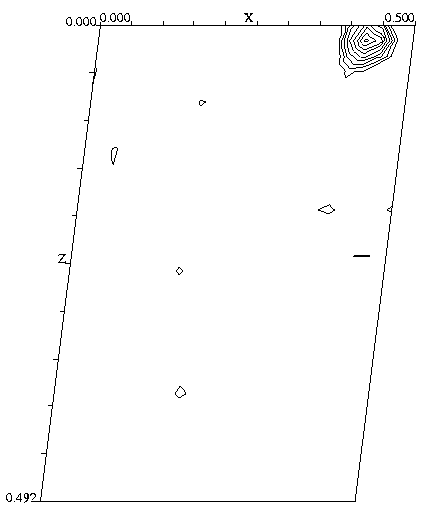
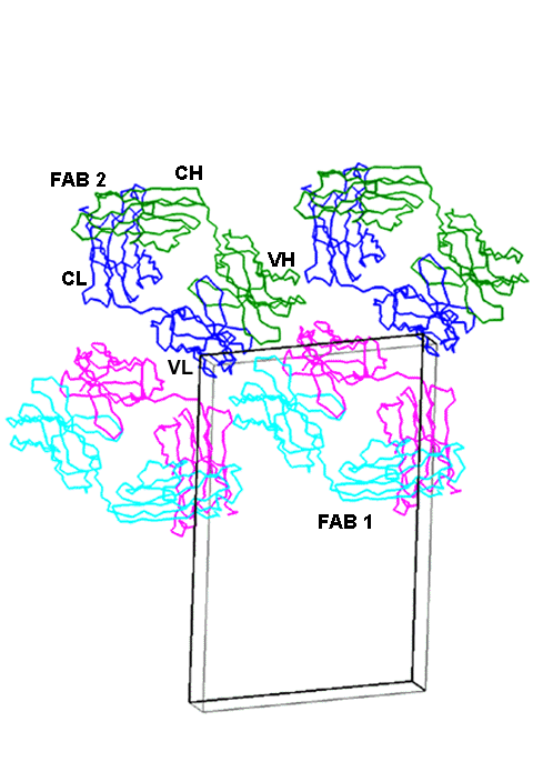
The structure has been solved by molecular replacement (MR) using murine Fab as a search model. Space group is P2(1) with 2 molecules in the asymmetric unit. A native patterson map reveals the position of the NCS symmetry axis parallel to the 2(1) axis. Click here for a view of the packing of Fab molecules in the 2E8 cell. FAB 1 and FAB 2 are NCS related through the 2-fold NCS axis located between the CDR3H contacts (green and light blue). The two other molecules are generated by crystallographic translation. Another NCS axis generated by crystallographic symmetry relates molecules FAB 1 and the one above on the right side.
![]() Back to X-ray Facility Introduction
Back to X-ray Facility Introduction
LLNL Disclaimer
This World Wide Web site conceived and maintained by
Bernhard Rupp (br@llnl.gov)
Last revised June 02, 2000 13:17
UCRL-MI-125269