 Our X-ray
Diffraction EquipmentOur X-ray
Diffraction Equipment
Our X-ray
Diffraction EquipmentOur X-ray
Diffraction Equipment
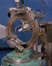
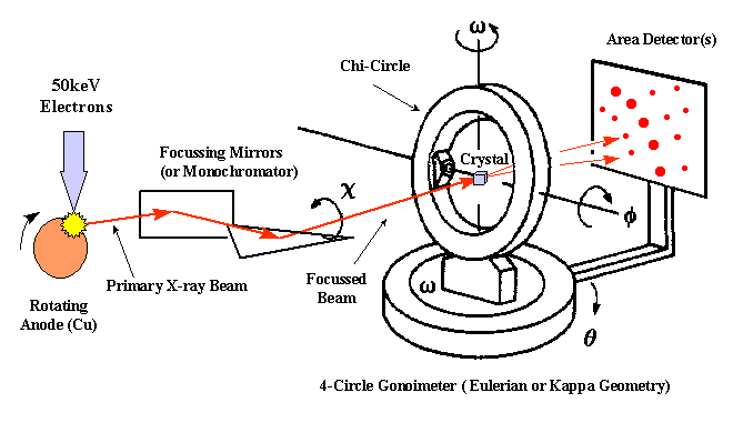
The Experimental Setup
To perform a X-ray diffraction experiment, we need an x-ray source. In most cases a rotating anode generator producing a X-ray beam of a characteristic wavelength is used. Intense, tunable X-ray radiation produced by a Synchrotron provides additional advantages. The primary X-ray beam is monochromated by either crystal monochromators or focusing mirrors. After the beam passes through a helium flushed collimator it passes through the crystal mounted on a pin on a goniometer head. The head is mounted to a goniometer which allows to position the crystal in different orientations in the beam. The diffracted X-rays are recorded using image plates, Multiwire detectors or CCD cameras.
Flash cooling protein crystals to cryogenic temperatures (~100 K) offers many advantages, the most significant
of which is the elimination of radiation damage. A part of the X-rays passing through the
crystal is scattered in different directions into a detector. In our facility we use
multiwire detectors and CCD detectors. They all deliver an
image of the diffraction spots. A large number of these images recorded from different
crystal orientations are processed (scaled and merged) into a final list of indexed
reflection intensities.
To learn more about cryo-cooling of crystals click on the picture 
![]() Back to X-ray Tutorial Introduction
Back to X-ray Tutorial Introduction
LLNL Disclaimer
This World Wide Web site conceived and maintained by
Bernhard Rupp (br@llnl.gov)
Last revised July 13, 2001 08:49
UCRL-MI-125269