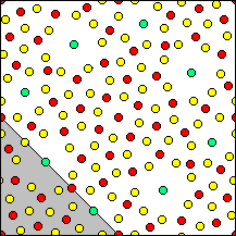
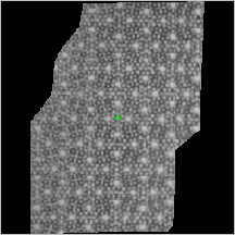
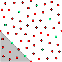
The final structure model and the corresponding Fourier map at 3Å resolution are plotted for one unit cell according to the results of J.J. Hu, F.H. Li and H.F. Fan, Ultramicroscopy, 41 (1992) 387-397. In the model, red balls represent Nb atoms, green balls represent K atoms, while yellow balls represent O atoms. The asymmetric unit at the lower-left corner is marked with gray color on the model and with green color on the Fourier map.
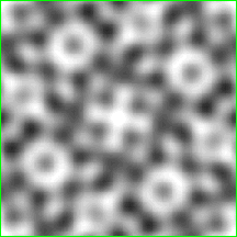
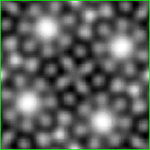
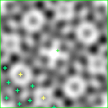
1. The original image and the average unit cell
![]()
![]()
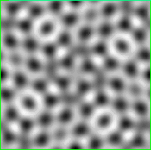
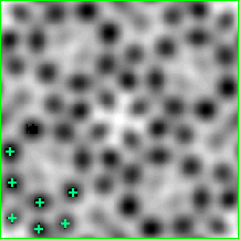
2. The first partial-structure model was derived from the deconvoluted image. This model contains 7 of the total 9 independent Nb atoms. Neither K atoms nor O atoms are included.
![]()
![]()
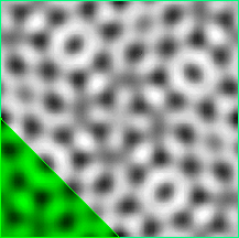
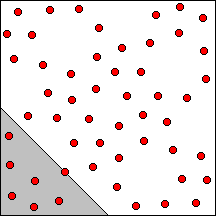
3 The second model was constructed from the Fourier map phased with the first model. This model contains all metal atoms except one Nb atom at the origin of the unit cell.
![]()
![]()
4. Fourier map (left) from the second model is compared with the final Fourier map at 3Å resolution (right). As is seen the Fourier map on the left resembles the final Fourier map in that, it possesses the feature of having four centered heptagonal channels surrounding by hexagonal, pentagonal, tetragonal and trigonal channels. It is concluded that the deconvolution of the single EM (far from Scherzer focus) and the subsequent modeling and Fourier recycling were successful. The missing of Nb atom at the unit-cell center can be explained by the low resolution limit of the deconvoluted data, since even the final 3 Å image (on the right part of the following figure) according to Hu et al. (1992) failed to reveal that Nb atom.